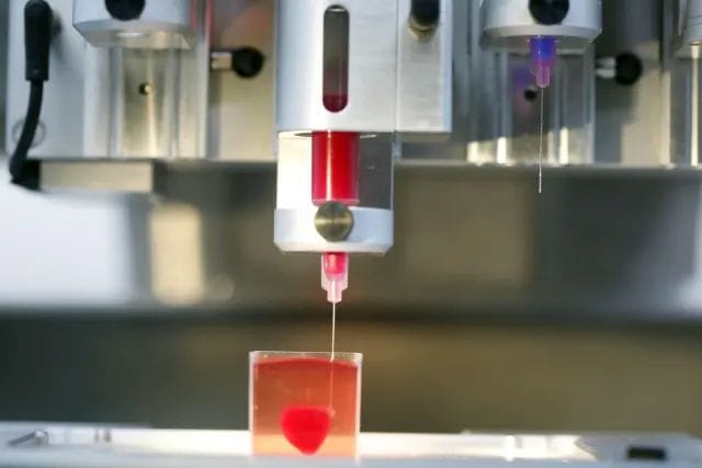
![3-D-printed heart engineered by Tel Aviv University scientists. [Source: Tomer Applebaum]](https://fabbaloo.com/wp-content/uploads/2020/05/image-asset_img_5eb09196618ab.jpg)
I’m reading a paper describing work done by Israeli scientists to literally 3D print a heart.
We first reported on this development back in April, and were very impressed with the progress in bioprinting. However, many news outlets are reporting this as the “world’s first 3D printed functional heart!” This is, of course, nonsense. The object produced by the Israeli scientists is not at all functional.
I’d like to clear the air about this project to counteract the mass media hype, as something like this seems irresistible to desperate reporters. It also gives me something to refer to when people will inevitably ask me about the “3D printed heart” when they see these reports.
That all said, the research does seem to be a rather large step forward. But I’d say it resembles more of a “world’s first 3D printed heart mock-up” than an actual functioning human organ.
Their approach used bioprinting, something we’ve written on many times. This branch of 3D printing involves using a syringe-based extrusion of living cells, typically onto a dissolvable scaffold. The living cells grow and eventually overrun the scaffold to form the desired biological tissue geometry.
Bioprinting Blood Vessels
The biggest problem facing bioprinting has been vascularization. This is the ability to produce blood vessels that carry nutrients to the living cells and carry away waste. Earlier bioprinting concepts simply dipped the 3D printed scaffold and tissue into a nutrient solution and let the tissue absorb what it could from the adjacent solution.
This approach works very well for thin biostructures, but is fatal for anything thicker. That’s because the nutrients cannot seep through to reach deeper cells. You need blood vessels to carry nutrients to them.
If you can imagine the step in complexity here, we would need to transform from essentially a mono-material uniform print to a differentiated multi-material, complex geometry print. In other words, add blood vessels to the structure.
3D Printing Stem Cells
![3D printed heart development concept [Source: Wiley]](https://fabbaloo.com/wp-content/uploads/2020/05/image-asset_img_5eb09196c5e99.jpg)
That is what the researchers really accomplished. They used skin cells that had been transformed into stem cells, which can generate any tissue. These they coaxed into two types of cells: heart cells for the muscle and epithelial for the blood vessels.
Then the really interesting (to me) part: they also devised software to generate a 3D model for the heart object that placed the blood vessels sufficiently close to all cells of the structure. To do this they used CT scans of actual hearts to develop a kind of “oxygen profile”.
![3D printed heart oxygen profile [Source: Wiley]](https://fabbaloo.com/wp-content/uploads/2020/05/image-asset_img_5eb0919715710.jpg)
They then 3D printed the heart and placed it in conditions mimicking the human body, which I presume would be circulating nutrients. Evidently, the cells did not die and properly reproduced, forming a heart-like structure.
Electric Shock to 3D Printed Heart
Even more interesting is the next step: the researchers introduced an electric shock and the cells began to contract!
However, before you get too excited, they were contracting randomly and individually; there was no coordination between them, and thus the “heart” was really incapable of pumping anything.
That’s why I’m terming this a kind of “mock-up” heart. It looks like one and has some similar characteristics, but isn’t really one. If you see those stories on CNN and other places, this is what it really is about.
An Actual 3D Printed Heart That Works?
To do the coordination I suspect the researchers would have to add more complexity to the print, specifically nerves to coordinate activities across the entire structure. That sounds complicated, but then so was the introduction of blood vessels. Perhaps we will see this as the next step?


FELIXprinters has released a new bioprinter, the FELIX BIOprinter, which is quite a change for the long-time 3D printer manufacturer.