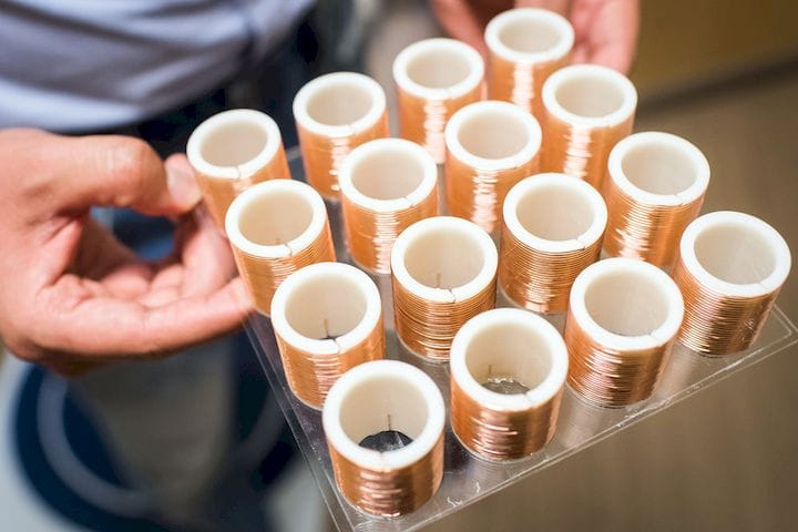
![A new magnetic metamaterial has been developed [Source: Boston University]](https://fabbaloo.com/wp-content/uploads/2020/05/image-asset_img_5eb095897b822.jpg)
Researchers at Boston University have developed a powerful new application of 3D printed magnetic metamaterials.
The problem being solved here is a common dilemma facing MRI operators. MRI machines use a form of magnetic resonance imaging to develop highly detailed 3D images of patient internals. These images are used by surgeons and others to diagnose medical issues or plan surgeries by “looking inside” the patient.
The problem is that in order to achieve sufficient detail, the magnetic field must be quite strong. In most MRI machines, the field is so powerful that any metal objects must be removed from the vicinity, lest they fly across the room and stick to the magnet. Even with this magnetic power harnessed, the images are not of the finest resolution and take considerable time to capture, often causing difficulties with patients who don’t appreciate being stuck inside a weird machine for long periods.
But if the magnetic field were cranked up to higher levels to achieve better resolution and speed captures, then larger areas would have to be cleared of metals, which is not good.
The new material is a kind of array of elements that each provide a platform for focusing and amplifying the magnetic field on a selective basis. We spoke with one of the lead researchers at BU about the project.
![Boston University researchers Xiaoguang Zhao, Xin Zhang, and Stephan Anderson [Source: Boston University]](https://fabbaloo.com/wp-content/uploads/2020/05/image-asset_img_5eb09589c7a7e.jpg)
Fabbaloo: Can you provide some details on the nature of this array? How large are the elements? Are there smaller components involved? How large is the array in total?
Xin Zhang: “The elements are individual helices with a diameter of 3 cm and height of 3 cm. The thickness of the copper wire is 0.28 mm. While this may seem large, when compared with the 5-meter RF wavelength that the metamaterial is operating at, the element is deeply sub-wavelength.
In addition, we can further minimize the element size by varying the design of the elements, which is one future direction of our research. There are no additional, smaller components involved in the elements.”
Fabbaloo: I’m not clear on how this works physically. In the video the array appears to be a slab that sits under the patient? How is the array actually positioned?
Xin Zhang: “We have flexibility to use this metamaterial physically. Options include utilizing a large array of helical resonators to form a slab upon which the patient is placed. On the other hand, for example, smaller, flexible arrays may be employed to conform to a kneecap. A variety of metamaterial designs may be employed for different imaging applications.”
3D Printed Metamaterial
Fabbaloo: How does the device do its job? Is it concentrating the magnetic field on certain areas, thus increasing the signal to noise ratio? Does this concentration cause potential issues for the patient?
Xin Zhang: “Simply speaking, there are two steps, a transmission phase followed by a reception phase. In the transmission phase, the coil in the MRI emits a radiofrequency (RF) magnetic field to excite the patient’s body. Next, the MRI coil listens to the magnetic field ‘radiated’ by the patient to form images of the patient.
Our metamaterial interacts with the magnetic field in both the transmission phase and reception phase. You are correct, the metamaterial concentrates the magnetic field in certain areas to increase the signal-to-noise ratio.
Utilizing our approach, this concentration does not cause potential issues for the patient. We decreased the RF power in the transmission phase to ensure that the subject to be imaging would be properly excited. In addition, our metamaterial does not exhibit electric field confinement, which would be of safety concern due to the potential for heating in the patient, as detailed in the paper and the supplemental information of the paper. Therefore, our magnetic metamaterial is likely to be safely translated towards human imaging.”
Fabbaloo: Are you 3D printing all of this together, or installing the copper after printing? What 3D printing technology/vendor/machine are you using?
Xin Zhang: “We print the scaffold of the metamaterial, which is the hollow cylinder featuring outer helical grooves, and then wound the copper wire around the scaffold. We employed the fused deposition modeling (FDM) technique to print the metamaterial. The machine we employed for printing was a uPrint/Dimension system from Stratasys.”
Metamaterial Medical Usage
Fabbaloo: What do you think the actual use of this technology would be in practical terms? Would medical agencies choose to increase the SN ratio of their images, or would they decrease the imaging time to get more patient throughput? Or both?
Xin Zhang: “The actual use of metamaterial in the clinical imaging is very promising. However, this will require a variety of clearances related to the safety. Using this technique, clinicians may choose to either increase the SNR of the images, increase resolution, or increase the throughput of imaging.”
This is one of many new metamaterials being developed as a direct result of 3D printing’s ability to create arbitrary geometries at small scales. It won’t be the last.
Via Boston University


A research thesis details the incredibly complex world of volumetric 3D printing. We review the highlights.