Performing surgery isn’t a perfect science, and even doctors need to practice their skills, especially when it comes to children.
To hone those skills, doctors practice various techniques using anatomical models or mannequins. However, these are typically limiting regarding complexity and feel, which limits the training.
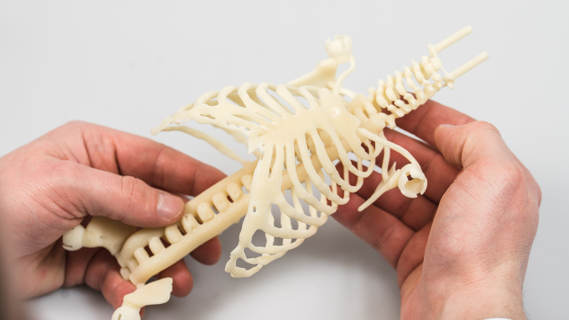
To overcome those limitations, Ph.D. grad student Mark Thielen (Eindhoven University of Technology) with the help of 3D Hubs, designed a shockingly detailed child mannequin that features a 3D printed skeleton with functional internal organs.
Mark’s prototype mannequin was designed using an MRI scan of an actual child to get the most accurate details.
Mark began the design of the model prototype using MRI scans he garnered from an actual infant, which was ideal for the level of detail needed for the project. He then enlisted the help of 3D Hubs to transition those 3D scans into an anatomically correct model and began testing several materials to see what worked best for the medical application.

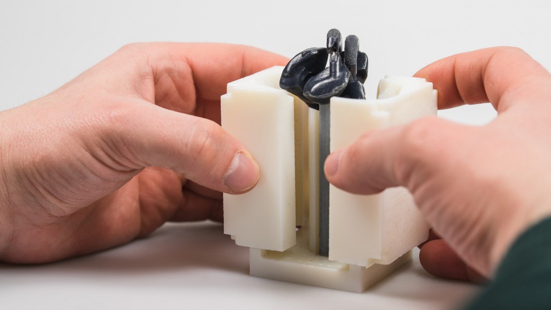
The model’s skeleton consists of two parts, the ribcage and spine, both 3D printed with an SLS 3D printer using thermoplastic elastomers, which house the functioning organs. Mark designed the heart and lungs to function similar to their organic counterparts. That is, the heart features working valves to pump fluid, and the lungs inflate/deflate to simulate breathing.
Read the rest at SolidSmack.com

