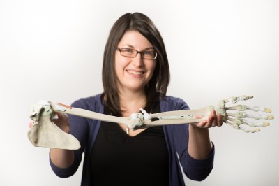
Medical students have learned anatomy and practiced surgery using cadavers for centuries. Now that doesn’t always have to be the case.
Researchers at Monash University in Melbourne, Australia have developed a commercially available “kit” containing “all the major parts of the body required to teach anatomy of the limbs, chest, abdomen, head and neck.”
Professor Paul McMenamin, Director of the University’s Centre for Human Anatomy Education, says:
For centuries cadavers bequested to medical schools have been used to teach students about human anatomy, a practice that continues today. However many medical schools report either a shortage of cadavers, or find their handling and storage too expensive as a result of strict regulations governing where cadavers can be dissected.
Without the ability to look inside the body and see the muscles, tendons, ligaments, and blood vessels, it’s incredibly hard for students to understand human anatomy. We believe our version, which looks just like the real thing, will make a huge difference.
Even when cadavers are available, they’re often in short supply, are expensive and they can smell a bit unpleasant because of the embalming process. As a result some people don’t feel that comfortable working with them.
Um, we’d agree with that.
The approach involves 3D scanning of the body components, then printing them out to the appropriate scale for use by students.
It’s one thing for such activities to be simulated by 3D software with images, but quite another for the topic to be physically displayed in a manner identical to reality. It seems clear that the same process can be used We can foresee this approach being used for many types of risky activities beyond medical training, including remote machine repair, planetary robotic activities and more.
Via PhysOrg

